There is clear evidence that light impacts our health and well-being. Besides the visual (image-forming) functions of the human ocular system, it is well known that it is also responsible for a number of non-visual functions, such as wake-sleep cycle, mood, alertness, and cognition, among others. [1-6] In fact, using these non-visual functions of the light, ocular photo-biomodulation (ocular PBM or ocular light therapy) has been used for decades to treat certain disorders, such as seasonal affective disorders (SADs), sleep problems, and circadian rhythm disruption. These biological and behavioral effects of light are enabled mainly by the melanopsin-containing intrinsically photosensitive retinal ganglion cells (ipRGCs) in addition to the conventional photoreceptors, rods and cones, present in the eye retina. [7-15] Ocular PBM has been also proven to have substantial potential benefits in treating various eye disorders, including that of myopia progression. [16-26] There is every reason to also believe that ocular light therapy, if applied with the proper intensity, modulation and wavelength of light, may provide substantial benefits in treating certain neurological disorders.
The challenge when using ocular light therapy has been to assure the light intensity, wavelength and repeated exposure of the ocular light therapy does not damage the eye’s fovea and macula. NeuroRays is pleased to announce Ocular Light Therapy with Macula Protection. The NeuroRays Ocular Light Therapy System represents the world’s first system capable of protecting the macula while applying ocular light therapy. The optics of the system have been designed using Zemax and the Liou Brennan eye model (Figure 1 below).

The proprietary system utilizes a sized and positioned fixation target that imparts a protective image over the macula and/or fovea when ocular light therapy is being applied. This protective image acts as a shield that protects potentially damaging wavelengths of light from striking the macula and/or fovea. This shield can be adjusted in diameter and degree of intensity and transmission of light. It can be of any color including black or a light filter. The shield can be adjusted to allow for 100% transmission to that of zero transmission.
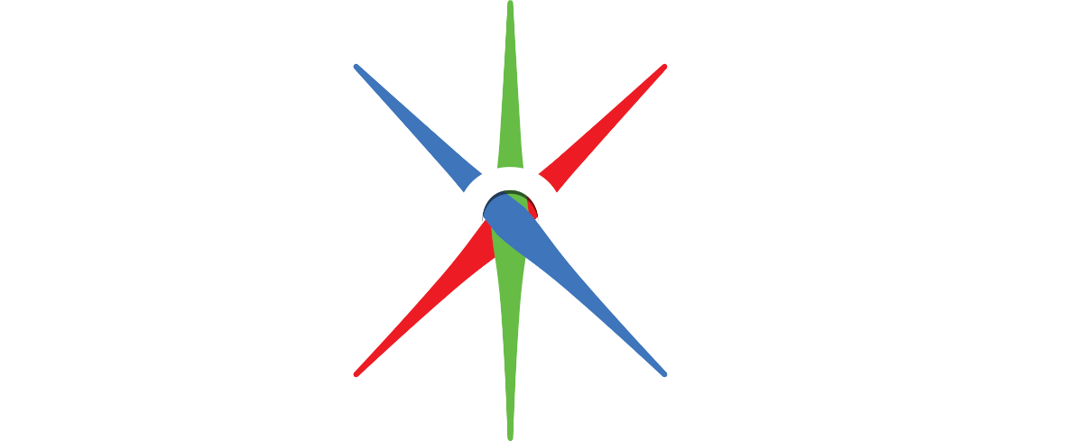
To better understand the system, think of the eclipse of the Sun for a moment. In a similar manner, NeuroRays’ proprietary fixation target, which is aligned with the eye’s line of sight, acts to eclipse the ocular light therapy light source. The eclipse casts a shadow like, protective image across the macula and/or fovea of the eye receiving ocular light therapy. This occurs while the visual aurora around the eclipse transitions from the full eclipse of light to that of the full surrounding ocular light therapy intensity and wavelengths that strike the retina peripheral to the macula and/or fovea (Figure 2).

This technical breakthrough allows for increasing the intensity of the ocular light source while protecting the macula and fovea. The ocular light can be of any wavelength desired for treating an eye disease or neurological disorder, like blue light therapy (BL) used for SAD-treatment, or repeated low-level red-light (RL) therapy for treating myopia. [7-26] The proprietary system can be designed to protect part or all of the macula and/or the fovea.
When utilizing the system for treating myopia progression the system can be used with or without myopia control lenses. The use of DIMS with ocular light therapy has recently shown encouraging results. Two recent third party independent clinical studies; one utilizing BL therapy in 2024 (which has not been published yet), and a 2025 published study utilizing red-light therapy has shown that the use of DIMS with ocular light therapy achieved 100% stoppage of myopia progression. [26] In addition, it has been shown that perfusion of the choroid (the thickening of the choroid) at the posterior pole of the eye occurs even when the retina is stimulated with ocular light therapy ~4.0 mm to ~5.0 mm from the macula.
While the ocular PBM (or ocular light therapy) has been used for decades, and it is generally accepted that the “low” level light has no acute impact on the eye, it is of vital importance to understand how the repeated light exposures impact the eye health, as well as the overall health and well-being in long terms. There are still debates on the potential toxicity of cumulative exposures of these light therapy treatments. [7-26]
For instance, bright light and blue light (RL) therapy have been used to treat SAD, to reset circadian rhythms, etc. [14, 15] Blue light exposure depending on the wavelength or intensity, may cause photo-chemical reactions in some of the eye tissues, especially the retina. The real toxicity of long-term blue light cumulative exposure and the dose dependent effect are currently unknown. However, it is well known that bad blue light wavelengths within the range of 400nm – 440nm can cause the most damage. The so-called blue light hazard (BLH) weighting function is widely used when considering the photochemical risk to the retinal tissues. BLH is defined over the 300–700 nm range with a peak ~ 440 nm (ICNIRP guidelines – International Commission on Non-Ionizing Radiation Protection, 2013). Therefore, BLH is wavelength-dependent, peaking in the range 430 nm – 440 nm, which is considered as the highest risk of photochemical injury for the retina. Please, note that the BLH range is distinct from the wavelength range responsible for non-image-forming light effects peaking at around 480 nm – 490nm (melanopsin-sensitivity curve). Moreover, it is not sufficient to know just the BLH-range, but also the prolonged and/or repeated exposure from focused (collimated) light sources. [27-31]
Another example is the red light (RL) therapy being used for treating various ocular and especially retinal diseases, such as myopia, retinal degeneration, diabetic retinopathy, retinopathy of prematurity, age-related macular degeneration, diabetic macular edema, retinitis pigmentosa, etc. [19-26, 32] However, the thermal and photochemical effects of RL are not fully understood. The retina (more specifically the macula) is at risk for photochemical and thermal damage when using repeated RL therapy, even when used at very low-level intensities. A recent study confirmed that kids receiving RLRL therapy showed lower cone density in the paracentral fovea compared with nonusers, with some participants displaying subtle retinal abnormalities. [33, 34]
Therefore, it is of paramount importance to understand how the light in these various ocular PBM (light) therapies is being received by different eye parts, once it enters the eye. Many questions and uncertainties, like those presented below, are still topics for debates:
- Where will the light beam hit once entering through the pupil? Will it damage the photoreceptors and other eye tissues?
- Which part of the retina will receive most of the light and at what intensity? Is it the fovea, the macula, or the whole retina?
- How the light is being administered by the light therapy source – as diffuse or as focused beam?
- Although some of the commercial PBM devices claim low intensity light, repeatedly applying it few times daily over the time period of year or longer, is a huge concern due to its accumulative effects – in fact, the macula sees a lot of light over a period of time.
- Obviously, light intensity plays a huge role; low-level light would require a longer therapy time period. Higher light intensity would work much better, shortening the exposure times, and the overall therapy time, but the problem is that higher intensity can be detrimental for the fovea and macula.
- Moreover, the tested subjects (e.g. kids with myopia) using RL therapy do not wear their glasses during the therapy, so the collimated light beam hits their fovea (or macula) and is not being diffused over the whole retina.
Thus, the concerns of light exposure during ocular PBM therapies, either being it with blue light, red light, or any other light wavelengths, or their combination, is understandable and should not be neglected.
Considering all the above concerns currently present in ocular PBM therapies, NeuroRays has conceived of, invented, proven with optical modeling, and patent protected the world’s first ocular light therapy system that enables the safe use of light beams of higher intensity. Using higher intensity light can lead to shorter exposure times thus resulting in hopefully shorter therapy periods. The NeuroRays approach in treating myopia (and other ocular and neurological disorders) with light, utilizes a novel sized and positioned fixation target that casts a protective image over part or all of the macula, regardless of the type of beam the light source is producing (collimated or not). While the protective image from the fixation target protects the macula, the rest of the retina can be exposed to the ocular therapy light wavelengths, thus allowing for the retina peripheral to the macula to receive most of the therapy light. For instance, rods and ipRGCs have been identified to be responsible for stopping myopia progression [35-37]; therefore, ocular light therapy exposure to the retinal periphery, where the most rods and part of the ipRGCs are located is the best-case scenario for treating myopia. This occurs while the macula remains protected even when higher intensity therapy light is applied. And by using myopia control lenses such as DIMS during ocular light therapy adds more to the spread of the therapy light over the retinal periphery. As stated earlier it has been shown that utilizing ocular light therapy striking the retina 5 mm away from the macula can elicit a thickening of the choroid at the posterior pole of the eye.
Furthermore, NeuroRays’ patent-protected approach also covers the use of safe blue light wavelengths. For instance, longer wavelength blue light therapy with wavelengths within the range (480 nm – 500nm) has been shown to activate both ipRGCs and rods, while avoiding the photo-chemical damage of ocular tissues as seen with shorter blue light wavelengths. These longer blue light wavelengths have been identified to stop the axial length of myopic eye and thicken the choroid. It is important to point out that the NeuroRays’ Ocular Light Therapy System allows for the use of most light wavelengths while providing protection to the macula. It is up to the operator to choose the level of macula protection, wavelength, intensity, and time of therapy based upon the eye or neurological disorder to be treated. The NeuroRays’ Ocular Light Therapy System can be incorporated into equipment and eyewear.
References
- Morioka, H.; Ozawa, H.; Kato, T. Physiological Study of Visual and Non-Visual Effects of Light Exposure. Appl. Sci. 2023, 13, 5785.
- Campbell, I. et al., Light as a Modulator of Non-Image-Forming Brain Functions—Positive and Negative Impacts of Increasing Light Availability. locks&Sleep 2023, 5, 116–140.
- Maruani, J. et al. Multi-Level Processes and Retina–Brain Pathways of Photic Regulation of Mood. J. Clin. Med. 2022, 11, 448.
- Wahl, S. et al., The inner clock—Blue light sets the human rhythm, J. Biophotonics. 2019; 12: e201900102
- Blume, C. et al., Effects of light on human circadian rhythms, sleep and mood, Somnologie 2019 · 23:147–156
- Vandewalle, G. et al., Light as a Modulator of Cognitive Brain Function, Trends in Cognitive Sciences, 2009, Vol.13 No.10
- Huang, X. et al., A Comprehensive Overview of the Neural Mechanisms of Light Therapy, Neurosci. Bull. March 2024, 40(3):350–362
- Vandewalle, G. et al., Spectral quality of light modulates emotional brain responses in humans, PNAS, 2010, vol. 107, no. 45 | 19549–19554
- Sun, W. et al., Efficacy and Safety of Light Therapy as a Home Treatment for Motor and Non-Motor Symptoms of Parkinson Disease: A Meta-Analysis, Med Sci Monit, 2022; 28: e935074
- Postolache, T.T. et al., Circadian Phase Shifting, Alerting, and Antidepressant Effects of Bright Light Treatment, Clin Sports Med 24 (2005) 381– 413
- Fernandez, D.C. et al., Light Affects Mood and Learning through Distinct Retina-Brain Pathways, Cell 175, 71–84, September 20, 2018
- Xhen, Z-W. et al., Treatment measures for seasonal affective disorder: A network meta-analysis, Journal of Affective Disorders 350 (2024) 531–536
- Alkozei A, et al., Exposure to Blue Wavelength Light Is Associated with Increases in Bidirectional Amygdala-DLPFC Connectivity at Rest. Front. Neurol. 2021, 12:625443.
- Meesters Y. et al., The effects of low-intensity narrow-band blue-light treatment compared to bright white-light treatment in sub-syndromal seasonal affective disorder, BMC Psychiatry (2016) 16:27
- Campbell, P.D. et al., Bright Light Therapy: Seasonal Affective Disorder and Beyond, Einstein J Biol Med. 2017; 32: E13–E25.
- Wang, X. et al., Effects of blue light exposure on ocular parameters and choroidal blood perfusion in Guinea pig, Experimental Eye Research 235 (2023) 109619
- Thakur, S. et al., Short-Term Exposure to Blue Light Shows an Inhibitory Effect on Axial Elongation in Human Eyes Independent of Defocus, IOVS | 2021 | Vol.62 | No.15 | Article 22
- Chakraborty, R. et al., Axial length reduction and choroidal thickening with short-term exposure to cyan light in human subjects, Ophthalmic Physiol Opt. 2024;00:1–19.
- Liu, Z. et al., The Effects of Repeated Low-Level Red-Light Therapy on the Structure and Vasculature of the Choroid and Retina in Children with Premyopia, Ophthalmol Ther (2024) 13:739–759
- Salzano, A.L. et al., Repeated Low-level Red-light Therapy: The Next Wave in Myopia Management? Optom Vis Sci 2023; 100:812–822.
- Xu, Y. et al., Repeated Low-Level Red-Light Therapy for Myopia Control in High Myopia Children and Adolescents, Ophthalmology 2024;131:1314-1323
- Gu, L. et al., Axial Shortening Effects of Repeated Low-level Red-light Therapy in Children with High Myopia: A Multicenter Randomized Controlled Trial, Am. J. Ophthalmology, 2025, 270, p203-215
- Jiang, Y. et al., Effect of Repeated Low-Level Red-Light Therapy for Myopia Control in Children – A Multicenter Randomized Controlled Trial, Ophthalmology 2022;129:509-519.
- He, X. et al., Effect of Repeated Low-level Red Light on Myopia Prevention Among Children in China With Premyopia, A Randomized Clinical Trial, JAMA Network Open. 2023; 6(4):e239612.
- Dong, J. et al., Myopia Control Effect of Repeated Low-Level Red-Light Therapy in Chinese Children, A Randomized, Double-Blind, Controlled Clinical Trial, Ophthalmology 2023;130:198-204.
- Yang, Y. et al., Synergistic effect of defocus incorporated multiple segment glasses and repeated low-level red-light therapy against myopia progression, Scientific Reports, (2025) 15:3996
- Wong, A. N. et al., A review of the current state of research on artificial blue light safety as it applies to digital devices, Heliyon 8 (2022) e10282
- Cougnard-Gregoire, A. et al., Blue Light Exposure: Ocular Hazards and Prevention— A Narrative Review, Ophthalmol Ther (2023) 12:755–788
- Ouyang, X. et al., Mechanisms of blue light-induced eye hazard and protective measures: a review, Biomedicine & Pharmacotherapy Volume 130, October 2020, 110577
- Chakravarthy H, Georgyev V, Wagen C, Hosseini A and Matsubara J (2024) Blue light-induced phototoxicity in retinal cells: implications in age-related macular degeneration. Front. Aging Neurosci. 16:1509434.
- Françon, A. et al., The blue light hazard and its use on the evaluation of photochemical risk for domestic lighting. An in vivo study, Environment International Volume 184, February 2024, 108471
- Zhang, C.-X.; Lou, Y.; Chi, J.; Bao, X.-L.; Fan, B.; Li, G.-Y. Considerations for the Use of Photobiomodulation in the Treatment of Retinal Diseases. Biomolecules 2022, 12, 1811.
- Liao, X. et al., Cone Density Changes After Repeated Low-Level Red-Light Treatment in Children with Myopia, JAMA Ophthalmol.2025
- Ostrin, L. A. et al., Red Light Instruments for Myopia Exceed Safety Limits, Ophthalmic Physiol Opt. 2024 March; 44(2): 241–248
- Perez-Fernandez, V. et al., Rod Photoreceptor Activation Alone Defines the Release of Dopamine in the Retina, 2019, Current Biology 29, 763–774, 2019.
- Liu, A-L. et al., The role of ipRGCs in ocular growth and myopia development, Sci. Adv. 8, eabm9027 (2022) 8
- Chakraborty, R. et al., Melanopsin modulates refractive development and myopia, Experimental Eye Research, 214, 2022, 108866